Outsourced to AI
  • About This Project
  • Tools Used in This Project
  • The Archive
Sign in Subscribe

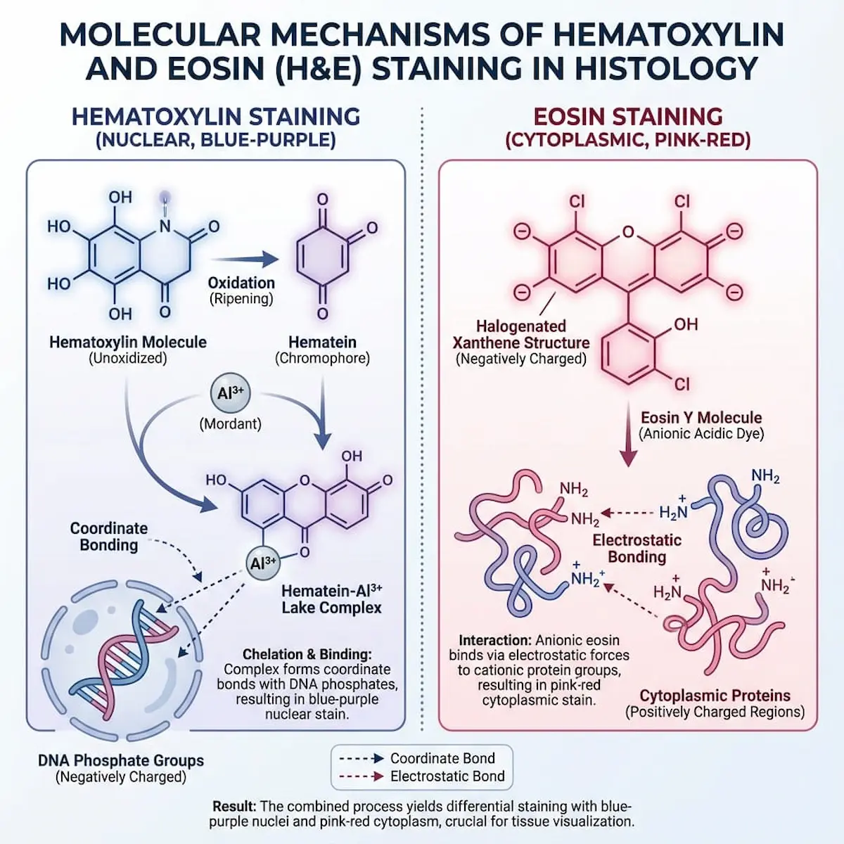
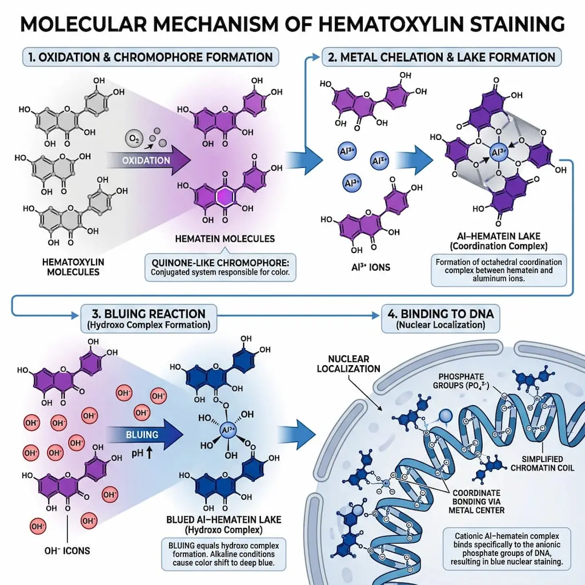
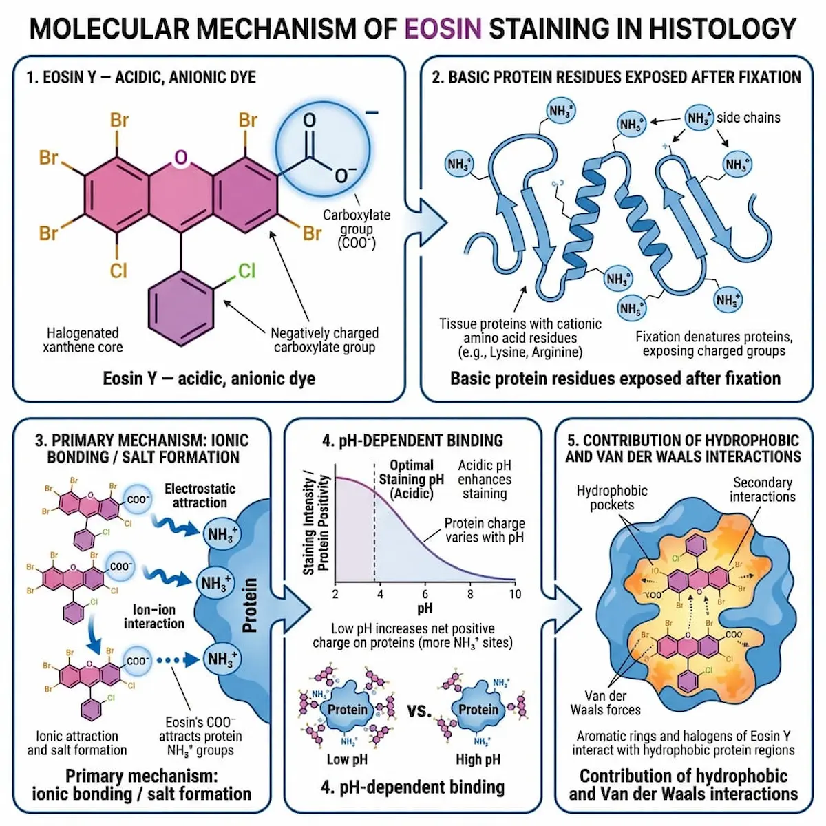
Hematoxylin and Eosin Infographic

Last updated on  22 May 2026
Infographic showing molecular mechanism of H&E staining: hematoxylin-aluminum binding to DNA phosphates, eosin binding electrostatically to proteins.
How H&E Staining Actually Works
Infographic showing hematoxylin staining mechanism in four steps: oxidation to hematein, aluminum chelation, bluing reaction, and DNA phosphate binding.
Four Steps Behind the Blue
Infographic of eosin staining mechanism in five panels: anionic dye structure, exposed protein residues, ionic bonding, pH dependence, hydrophobic forces.
Why Eosin Stains Pink
Previous AI in Histotechnology Infographic
Next Histotechnology in Drug Development
Outsourced to AI © 2026
Powered by Ghost