Protocol Transfer Is Not Copy and Paste
A familiar story plays out in laboratories everywhere. An assay performs beautifully in one place. The protocol gets written down, sent to a second lab, followed step by step, and the result comes back wrong: too weak, too strong, buried in background, or simply not interpretable. The people involved are competent. The steps were honored. And still the stain refuses to behave.
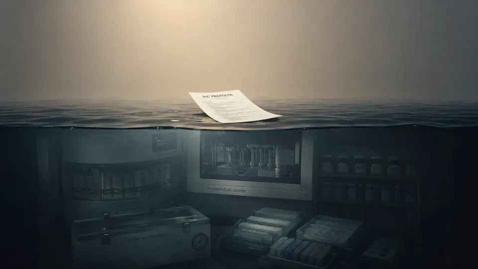
The reason is rarely incompetence. It is a misunderstanding about what a protocol actually is. We tend to treat the written method as the whole method, when in truth it is only the visible portion of something much larger. An IHC or histology protocol is the output of an entire laboratory environment, and when it travels, every hidden condition that shaped it stays behind unless someone deliberately carries it along.
The part people think they are sending
Pressed on what we are sending, most of us would name the obvious technical anchors: antibody clone, dilution, incubation time, retrieval condition, detection system, chromogen, counterstain, and a suitable control. None of that is wrong. Those details are necessary. They are also incomplete. A written sheet records what was done; it seldom captures how the surrounding lab quietly shaped the outcome. The protocol is partly technical and partly cultural. It encodes how tissue is handled, how a tech judges a section, how a pathologist reads a slide, and how problems get fixed when something looks off.
Failure often begins before staining
It is tempting to hunt for the fault inside the autostainer, but a transfer can break long before a slide reaches the instrument. Consider fixation. Two labs can both claim to fix in formalin while differing in time to fixation, total fixation duration, specimen size, formalin volume relative to tissue, and the age or quality of the formalin itself. Underfixation and overfixation pull staining in opposite directions, and no change to antibody concentration can repair tissue that arrived at the bench with a different fixation history.
In clinical work, that variation tends to originate in collection and grossing. In pharma, it surfaces through necropsy timing, study logistics, shipment, or how a contract lab handled the material. Processing adds another layer. Dehydration and clearing times, paraffin temperature, reagent upkeep, section thickness, and the processor model all influence morphology and antigen preservation. When the tissue entering the stain is not equivalent, the stain cannot be equivalent either, no matter how faithfully the steps are copied.
The same step is not always the same step
Even within the staining run, identical wording can hide real divergence. "High pH retrieval" sounds like a fixed instruction, yet the instrument, buffer formulation, exact pH, temperature, time at temperature, cooling profile, and whether retrieval happens under pressure, in a water bath, onboard, or offline can each move a result. Retrieval is doing real chemistry on the section, reversing crosslinks and exposing the target rather than simply heating the slide, so a modest shift in any of those parameters can carry staining from faint to optimal, or from clean to noisy.
Detection chemistry deserves the same scrutiny. Polymer systems vary in sensitivity, species cross-reactivity, and amplification behavior, and their compatibility with the primary antibody's host species matters as much as the antibody itself. This becomes acute in pharma, where a single study may juggle human tissue, mouse xenografts, and other animal models, each capable of generating its own background. Swap the kit and sensitivity, specificity, and how readable the slide turns out can all move with it.
Automation flattens none of this. Two autostainers can dispense reagents differently, heat slides differently, hold incubation environments differently, wash with different stringency, and run proprietary timing that no one outside the vendor fully controls. A method tuned to one platform rarely lands cleanly on another without adjustment.
Small variables compound
Beyond the headline parameters sit dozens of quiet ones. A fresh antibody or detection lot behaves a little differently from the last. Charged and uncharged slides grip sections unevenly. Then come the half micron of extra thickness, the few minutes of drying lost or gained, the older cut section, the harder water, the unfamiliar coverslipping medium, and the counterstain dialed a shade too strong. Any one of these reads as trivial. The danger is additive. A failed transfer is usually not a single dramatic error but a slow accumulation of minor mismatches that, stacked together, push the assay past the point where it still reads the way it used to.
Agreeing on what "working" means
A protocol can run flawlessly and still fail to transfer if the two labs disagree about what an acceptable slide looks like. People bring different thresholds for what counts as positive, how much background is tolerable, whether signal belongs at the membrane or in the cytoplasm, and how to separate weak true staining from nonspecific noise. Tumor versus stromal localization, a pathologist's personal threshold, and a biomarker team's expectations all factor in. The handoff is not finished when the chromogen develops. It is finished when the people who depend on the result interpret it the same way.
That endpoint shifts depending on where you stand. Clinical labs are built around reproducibility, validated performance, regulatory expectations under frameworks like CAP and CLIA, and readiness for diagnostic sign-out. A shaky transfer there means repeat testing, troubleshooting, delays, and potential risk to patient care. Pharma labs more often live with exploratory biomarkers, limited and precious tissue, multiple contract labs, evolving study questions, and assays that may have no established clinical cutoff yet. A failed transfer in that world rarely announces itself as a failed diagnostic. It shows up as confusing data, inconsistent staining, and a quiet erosion of confidence in the underlying biology.
What the paper never captures: judgment
Here is the part that resists documentation. A written protocol cannot, on its own, hand over the instinct for when to stop optimizing, when background has crossed from acceptable to disqualifying, when a control is lying to you, when the real problem is tissue quality rather than chemistry, when to pull in the pathologist, and when a method simply should not be forced onto a tissue type it was never built for. Much of that judgment lives at the bench, carried without ever being named. What breaks during transfer is frequently not the protocol at all, but the experience that surrounded it.
Transferring the method, not just the sheet
Better transfer treats the protocol as one item in a package rather than the package itself. A strong handoff carries the full method alongside platform specifics, antibody clone and lot, detection kit and lot, retrieval buffer and instrument, processing details, section thickness, slide type, and drying conditions. Control examples travel with it, along with the expected staining pattern, an acceptable background level, representative images, known failure modes, and the troubleshooting history the receiving lab would otherwise rediscover the hard way. The interpretation criteria come too, with a clear note on who signs off on them. For pharma, the package also names the species and model in play, flags xenograft-specific background concerns, says whether the assay is exploratory or tied to a decision, and marks any limits imposed by scarce tissue.
The vocabulary tends to shift with experience as well. "Use the same protocol" carries a quiet promise of identity, and labs that have been burned by it lean toward re-establishing the protocol under local conditions instead. They are slower to call an assay failed and quicker to note that the method has not yet reproduced under the receiving lab's pre-analytical, analytical, and interpretive conditions. What actually moves between two labs is the method, the materials, the controls, the expectations, and the judgment behind them, never the sheet alone.
Protocols live inside systems
A protocol does not work because it is written down. It works because many conditions happen to align at once. Tissue handling, the reagents and instruments in play, the controls anchoring the run, the way a section is cut and dried, and the eye that finally reads the slide all have to cooperate. Change any of those and the method may need to be requalified, reoptimized, or reinterpreted before it earns trust again.
So the honest frame is not copying but translation. Move a method between labs and you are moving it between two languages of practice. Get the words across without the context, and the meaning is exactly what gets lost.