β-Galactosidase as a Functional Readout in Drug Development
What β-Galactosidase Staining and IHC Can Answer That Other Assays Cannot
Abstract
β-Galactosidase (BGAL) staining has been widely used as a reporter of gene expression and cellular state in preclinical research. In drug development, however, its value extends beyond simple expression mapping. When applied correctly, BGAL staining serves as a spatially resolved, functional readout that integrates transcriptional activity, protein translation, enzymatic competence, and cellular microenvironment. This paper outlines the biological basis of BGAL staining, distinguishes enzymatic activity from expression, and frames the specific experimental questions BGAL can answer across discovery, preclinical validation, and translational decision making.
Introduction
Drug development increasingly relies on multi-layered biological evidence. Transcriptomics identifies what genes are being transcribed. Proteomics measures what proteins are present. Functional assays attempt to determine whether those molecular signals translate into meaningful biological activity.
β-Galactosidase staining occupies a distinct and often misunderstood position in this hierarchy. Rather than directly reporting transcription or protein abundance, BGAL staining reports functional enzymatic activity preserved at the time of tissue harvest. This distinction is critical when interpreting results in pharmacology, toxicology, and translational research.
Understanding what BGAL does and does not report allows it to be used intentionally rather than incidentally.
Biological Basis of β-Galactosidase Staining
From transcription to stain
The biological sequence underlying BGAL signal is:
mRNA transcription → protein translation → protein folding and stability → enzymatic activity → substrate conversion → visible precipitate
β-Galactosidase staining reflects only the final steps of this sequence. Signal is generated only when the enzyme is present, properly folded, catalytically active, and situated in an environment that permits substrate conversion.
As a result, BGAL staining is a downstream, integrative readout, not a direct measurement of expression.
BGAL Staining Versus BGAL IHC
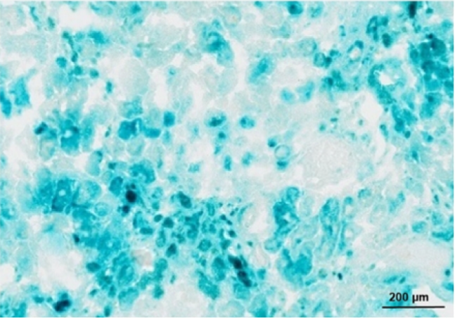
Enzymatic staining
Traditional BGAL staining, such as X-gal–based assays, requires preserved enzymatic activity and is therefore typically performed on frozen tissue or lightly fixed samples. The resulting signal reflects where the enzyme was active, not merely present.
BGAL IHC
BGAL immunohistochemistry detects the presence and localization of the β-galactosidase protein, regardless of enzymatic activity. It can be performed on paraffin-embedded tissue and aligns conceptually with other protein-level assays.
In drug development, these approaches answer different questions:
- BGAL staining answers whether a biological pathway is functionally engaged
- BGAL IHC answers whether the reporter protein is present and where it is localized
Used together, they separate protein presence from protein function.
Core Questions BGAL Can Answer in Drug Development
1. Is a target pathway functionally engaged in vivo?
In reporter models where lacZ is driven by a pathway-specific promoter, BGAL staining identifies tissues or cell populations in which the pathway is not only transcribed but functionally realized.
This is particularly valuable when:
- mRNA induction does not correlate with phenotypic response
- post-transcriptional regulation is suspected
- pathway activation is heterogeneous within tissue architecture
BGAL staining provides spatial confirmation that pathway engagement occurs where it matters biologically.
2. Does a drug modulate activity rather than expression?
Many compounds alter cellular function without producing proportional changes in mRNA levels. BGAL staining can reveal:
- suppression of functional activity despite unchanged transcript levels
- delayed or persistent activity after transcription has ceased
- regional differences in drug response within the same tissue
In this context, BGAL acts as a functional pharmacodynamic readout rather than a transcriptional biomarker.
3. Where does drug action succeed or fail within tissue microenvironments?
Bulk assays average signal across heterogeneous cell populations. BGAL staining preserves tissue context and can distinguish:
- tumor versus stroma
- epithelium versus immune infiltrate
- boundary regions where drug penetration or signaling breaks down
This spatial resolution is particularly important in oncology, fibrosis, and neurobiology.
4. Does functional activity persist after treatment withdrawal?
Because β-galactosidase protein can be stable, BGAL staining may persist after mRNA levels decline. This property allows investigators to identify:
- historical pathway activation
- durable biological effects of transient dosing
- regions of sustained functional change despite normalized transcription
In drug development, this helps differentiate temporary exposure effects from lasting biological impact.
5. Are observed transcriptional changes biologically meaningful?
High-throughput transcriptomic screens often identify numerous regulated genes. BGAL reporter systems can be used as a validation filter by asking:
- Does transcriptional induction translate into functional enzymatic activity?
- Are transcriptional changes biologically silent or functionally productive?
BGAL staining therefore helps prioritize targets that move beyond expression into action.
6. Are cells entering senescence as a treatment consequence?
In senescence-associated β-galactosidase assays, BGAL activity at acidic pH serves as a surrogate for lysosomal expansion and altered cellular metabolism. In drug development, this addresses questions such as:
- Does treatment induce senescence rather than apoptosis?
- Is senescence localized or widespread?
- Does senescence correlate with efficacy or toxicity?
This application is particularly relevant in oncology and aging-related therapeutic areas.
Relationship Between mRNA, BGAL IHC, and BGAL Staining
These assays should be viewed as complementary layers:
- mRNA assays report transcriptional intent
- BGAL IHC reports protein presence and localization
- BGAL staining reports functional enzymatic activity
Divergence between these layers is not experimental failure but biological information. A drug that suppresses BGAL activity without reducing mRNA reveals post-transcriptional or functional regulation. Conversely, persistent BGAL staining after mRNA decline reveals temporal lag and protein stability.
Limitations and Interpretive Boundaries
BGAL staining should not be used to:
- quantify expression levels precisely
- infer real-time transcriptional dynamics
- replace molecular assays in isolation
Its strength lies in contextualized function, not numerical precision.
Conclusion
β-Galactosidase staining and BGAL IHC occupy a unique niche in drug development. They answer questions that neither mRNA nor protein quantification alone can resolve. By reporting functional activity within intact tissue architecture, BGAL assays bridge the gap between molecular signal and biological consequence.
When used deliberately and interpreted correctly, BGAL becomes less a legacy reporter and more a strategic tool for functional validation, pharmacodynamic insight, and translational confidence.
Histotechnology Considerations for β-Galactosidase IHC and Enzymatic Staining
Successful interpretation of β-galactosidase data in drug development depends heavily on upstream histotechnology decisions. Unlike many conventional IHC targets, BGAL assays are unusually sensitive to tissue handling, fixation chemistry, and processing timelines. Failure to account for these factors can result in false negatives, misleading spatial patterns, or incorrect biological conclusions.
This section outlines practical considerations that determine whether BGAL staining reports biology or artifact.
1. Tissue handling and time to preservation
β-Galactosidase enzymatic activity begins to degrade immediately after tissue excision. Delays between harvest and preservation can lead to regional loss of signal that mimics biological heterogeneity.
Key considerations:
- Minimize ischemic time prior to freezing or fixation
- Standardize harvest-to-preservation intervals across treatment groups
- Avoid temperature fluctuations during transport
In drug studies comparing cohorts, inconsistent handling alone can appear as treatment effect.
2. Frozen versus fixed tissue decision point
The choice between enzymatic BGAL staining and BGAL IHC must be made before tissue processing.
Enzymatic BGAL staining:
- Requires frozen tissue or very mild fixation
- Preserves functional activity
- Is incompatible with routine paraffin workflows
BGAL IHC:
- Compatible with formalin-fixed paraffin-embedded tissue
- Detects protein presence rather than activity
- Is more forgiving for archiving and retrospective analysis
Histotechnical planning should align with the biological question, not convenience.
3. Fixation chemistry and duration
For BGAL IHC, fixation impacts epitope integrity rather than enzymatic activity. Overfixation can still reduce signal by masking epitopes.
Recommendations:
- Use neutral buffered formalin with controlled fixation times
- Avoid prolonged fixation beyond validated windows
- Validate antigen retrieval conditions specifically for BGAL
For enzymatic staining, even brief exposure to harsh fixatives can irreversibly inactivate the enzyme.
4. Section thickness and substrate penetration
β-Galactosidase enzymatic staining relies on substrate diffusion. Section thickness directly affects signal intensity and uniformity.
Typical considerations:
- Thinner sections improve substrate penetration but reduce total enzyme content
- Thicker sections may show surface-heavy staining with weak internal signal
- Consistency across slides is critical for comparative interpretation
Uneven staining across a section is often technical rather than biological.
5. Endogenous β-galactosidase background
Many tissues contain endogenous lysosomal β-galactosidase activity, particularly in:
- Macrophages
- Senescent cells
- Certain epithelial compartments
Histotechnical controls are essential:
- Include appropriate negative controls
- Use pH conditions specific to reporter versus senescence assays
- Interpret signal in anatomical context rather than color intensity alone
Failure to account for endogenous activity can lead to false attribution of reporter activation.
6. Antigen retrieval considerations for BGAL IHC
BGAL is a bacterial enzyme in most reporter systems. Antigen retrieval conditions optimized for mammalian proteins may not be optimal.
Practical guidance:
- Validate retrieval buffers empirically
- Avoid excessive heat that increases background
- Monitor tissue morphology alongside signal recovery
Strong signal with poor morphology is rarely acceptable in drug development settings.
7. Batch effects and longitudinal studies
BGAL staining is particularly vulnerable to batch variability.
Sources include:
- Substrate lot differences
- Incubation time drift
- Temperature variation
- Operator technique
In longitudinal drug studies, histotechnical standardization is critical to avoid confounding temporal effects with biological change.
8. Interpretation requires histology context
Histotechs play a key role in ensuring interpretable results by preserving:
- Tissue orientation
- Structural landmarks
- Compartmental integrity
BGAL signal without histological context loses much of its value. A technically perfect stain on poorly oriented tissue still limits biological insight.
Practical Implications for Drug Development Teams
For BGAL assays, histotechnology considerations are not downstream details. They are experimental variables that directly influence conclusions about drug efficacy, mechanism, and safety.
Early alignment between:
- biology teams
- pathology
- histotechnology
- translational science
reduces failed experiments, ambiguous data, and costly repetition.
Summary
β-Galactosidase assays demand more from histotechnology than many routine IHC targets. When tissue handling, fixation, sectioning, and controls are optimized, BGAL provides high-value functional insight. When they are not, BGAL quietly reports the condition of the workflow rather than the biology.
Comparative Overview of BGAL Enzymatic Staining, BGAL IHC, and RNAscope
A Histotechnology Perspective for Drug Development
|
Feature |
BGAL
Enzymatic Stain (X-gal) |
BGAL
IHC |
RNAscope |
|
What it
detects |
Functional
β-galactosidase enzymatic activity |
β-galactosidase
protein presence |
Target
mRNA transcripts |
|
Biological
layer reported |
Enzyme
activity (functional outcome) |
Protein
expression and localization |
Transcriptional
expression |
|
Primary
question answered |
Where is
the reporter biologically active? |
Where is
the reporter protein present? |
Where is
the gene being transcribed? |
|
Tissue
requirement |
Fresh
frozen or lightly fixed tissue |
FFPE or
frozen |
FFPE
(preferred), frozen possible |
|
Sensitivity
to tissue handling |
Very high |
Moderate |
High |
|
Tolerance
to fixation |
Poor |
Moderate
to good |
Very high
sensitivity to overfixation |
|
Fixation
constraints |
Minimal or
very short fixation only |
Requires
validated fixation window |
Strict
fixation time and chemistry required |
|
Compatibility
with paraffin |
No |
Yes |
Yes |
|
Antigen
or probe stability |
Enzyme
activity easily lost |
Protein
generally stable |
RNA highly
labile |
|
Section
thickness sensitivity |
High
(substrate penetration dependent) |
Moderate |
Moderate |
|
Background
risk |
Endogenous
β-gal activity |
Non-specific
antibody binding |
Low if
controls are correct |
|
Key
histotech failure mode |
False
negatives from enzyme inactivation |
Epitope
masking or retrieval failure |
RNA
degradation or overfixation |
|
Batch-to-batch
variability |
High |
Moderate |
Moderate
to high |
|
Spatial
resolution |
Good, but
enzymatic diffusion possible |
Good |
Excellent,
single-cell resolution |
|
Quantitative
potential |
Low |
Semi-quantitative |
High (dot
counting) |
|
Archival
suitability |
Poor |
Good |
Good |
|
Turnaround
time |
Short but
labor-sensitive |
Moderate |
Longer and
workflow-intensive |
|
Best
use case in drug development |
Functional
pathway engagement |
Protein
localization confirmation |
Transcriptional
validation |
|
Histotech
skill dependency |
Very high |
High |
Very high |
|
Reviewer
perception |
Functional
but contextual |
Standard
and accepted |
Gold
standard for spatial mRNA |
Practical Interpretation From the Histology Bench
- BGAL enzymatic staining is the most biologically integrative but the least forgiving. When it works, it answers questions no other assay can. When it fails, it often fails silently.
- BGAL IHC is the most practical bridge between discovery biology and translational pathology. It trades functional insight for robustness and archival compatibility.
- RNAscope provides unmatched transcriptional resolution but demands strict histotechnical discipline. Poor fixation or RNA handling can invalidate entire studies.
From a histotech standpoint, these assays should be viewed as complementary tools, not substitutes. Each one interrogates a different biological layer and places different demands on tissue handling and processing.
How this table is usually used in practice
- To justify why frozen tissue was required
- To explain discordant results across assays
- To guide endpoint selection during study design
- To educate non-histology stakeholders on why “just run it another way” is often not trivial
• • •